Pelvic Congestion Syndrome
It is estimated that one-third of all women will experience chronic pelvic pain in their lifetime. Many of these women are told the problem is “all in their head” but recent advancements now show the pain may be due to hard to detect varicose veins in the pelvis, known as pelvic congestion syndrome.
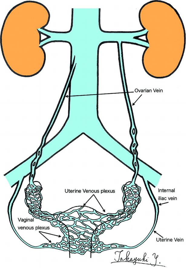
The causes of chronic pelvic pain are varied, but are often associated with the presence of ovarian and pelvic varicose veins. Pelvic congestion syndrome is similar to varicose veins in the legs. In both cases, the valves in the veins that help return blood to the heart against gravity become weakened and don’t close properly, this allows blood to flow backwards and pool in the vein causing pressure and bulging veins. In the pelvis, varicose veins can cause pain and affect the uterus, ovaries and vulva. Up to 15 percent of women, generally between the ages of 20 and 50, have varicose veins in the pelvis, although not all experience symptoms.
The diagnosis is often missed because women lie down for a pelvic exam, relieving pressure from the ovarian veins, so that the veins no longer bulge with blood as they do while a woman is standing.Many women with pelvic congestion syndrome spend many years trying to get an answer to why they have this chronic pelvic pain. Living with chronic pelvic pain is difficult and affects not only the woman directly, but also her interactions with her family, friends, and her general outlook on life. Because the cause of the pelvic pain is not diagnosed, no therapy is provided even though there is therapy available.
If you have pelvic pain that worsens throughout the day when standing, you may want to seek a second opinion with an interventional radiologist, who can work with your gynecologist. You can ask for a referral from your doctor, call the radiology department of any hospital and ask for interventional radiology or visit the doctor finder link at the top of this page to locate a doctor near you.


Prevalence
- Women with pelvic congestion syndrome are typically less than 45 years old and in their child bearing years.
- Ovarian veins increase in size related to previous pregnancies. Pelvic congestion syndrome is unusual in women who have not been pregnant.
- Chronic pelvic pain accounts for 15% of outpatient gynecologic visits.
- Studies show 30% of patients with chronic pelvic pain have pelvic congestion syndrome (PCS) as a sole cause of their pain and an additional 15% have PCS along with another pelvic pathology.
Risk Factors
- Two or more pregnancies and hormonal increases
- Varicose veins in thighs and groin
- Polycystic ovaries
- Hormonal dysfunction
Symptoms
The chronic pain that is associated with this disease is usually dull and aching. The pain is usually felt in the lower abdomen and lower back. The pain often increases during the following times:
- Pregnancy
- When tired or when standing (worse at end of day)
- Menstrual periods
- Following intercourse
Diagnosis and Assessment
Once other abnormalities or inflammation has been ruled out by a thorough pelvic exam, pelvic congestion syndrome can be diagnosed through several minimally invasive methods. An interventional radiologist, a doctor specially trained in performing minimally invasive treatments using imaging for guidance, will use the following imaging techniques to confirm pelvic varicose veins that could be causing chronic pain.
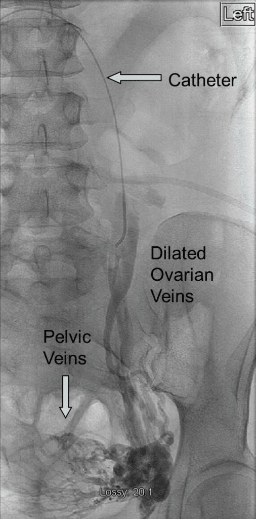
Pelvic Venography: Thought to be the most accurate method for diagnosis, a venogram is performed by injecting contract dye in the veins of the pelvic organs to make them visible during an X-ray. To help accuracy of diagnosis, interventional radiologists examine patients on an incline, because the veins decrease in size when a woman is lying flat.
MRI: May be the best non-invasive way of diagnosing pelvic congestion syndrome. The exam needs to be done in a way that is specifically adapted for looking at the pelvic blood vessels. A standard MRI may not show the abnormality.
Pelvic and Transvaginal Ultrasound: Not effective in the diagnosis of pelvic congestion syndrome as these exams are more focused on the uterus and ovaries and is performed lying flat. Although the varices can be visualized on ultrasound, the degree of dilation and number of veins is more difficult to grade than with venography or MRI.
Vertebroplasty is an outpatient procedure using X-ray imaging and conscious sedation. The interventional radiologist inserts a needle through a nick in the skin in the back, directing it under fluoroscopy (continuous, moving X-ray imaging) into the fractured vertebra. The physician then injects the medical-grade bone cement into the vertebra. Vertebroplasty takes from one to two hours to perform depending on how many bones are treated. The cement hardens within 15 minutes and stabilizes the fracture, like an internal cast.
Treatment Options
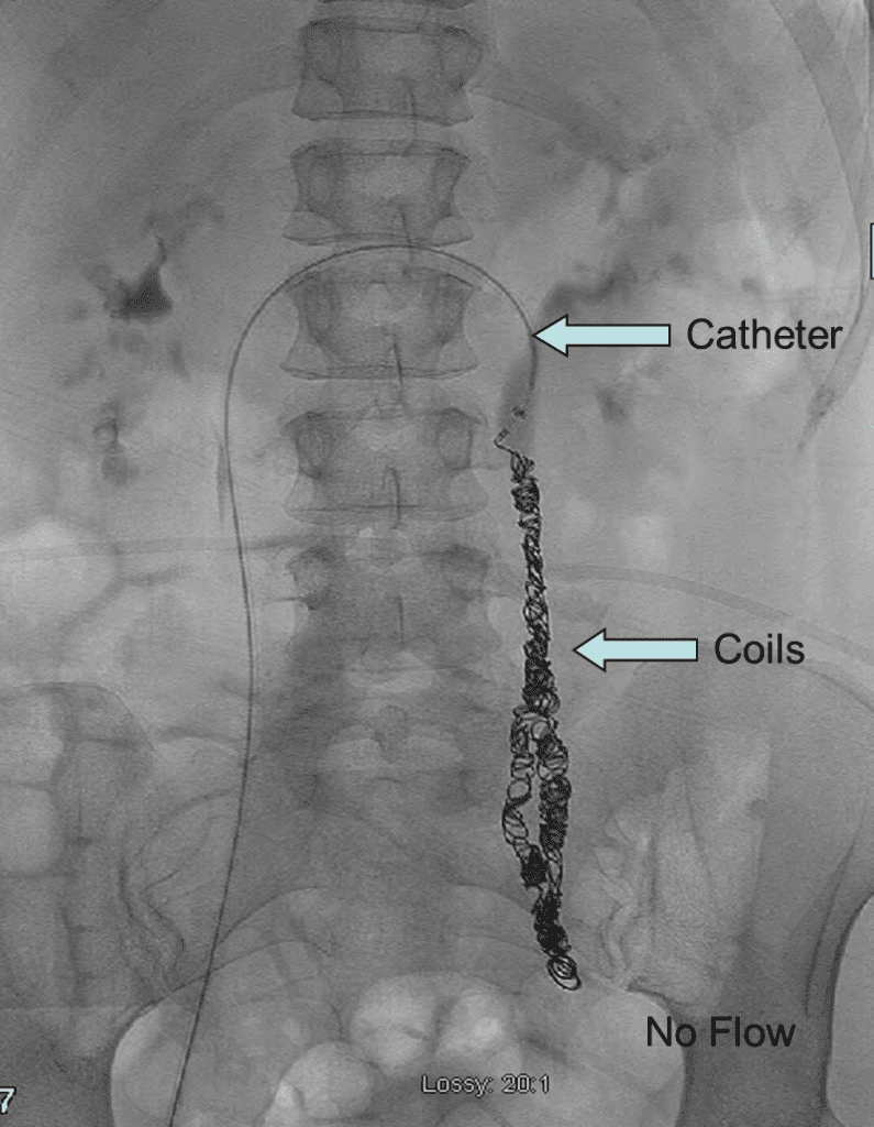
Once a diagnosis is made, if the patient is symptomatic, an embolization should be done. Embolization is a minimally invasive procedure performed by interventional radiologists using imaging for guidance. During the outpatient procedure, the interventional radiologist inserts a thin catheter, about the size of a strand of spaghetti, into the femoral vein in the groin and guides it to the affected vein using X-ray guidance. To seal the faulty, enlarged vein and relieve painful pressure, an interventional radiologist inserts tiny coils often with a sclerosing agent (the same type of material used to treat varicose veins) to close the vein. After treatment, patients can return to normal activities immediately.
Additional treatments are available depending on the severity of the woman’s symptoms. Analgesics may be prescribed to reduce the pain. Hormones such birth control pills decrease a woman’s hormone level causing menstruation to stop may be helpful in controlling her symptoms. Surgical options include a hysterectomy with removal of ovaries, and tying off or removing the veins.
Efficacy
In addition to being less expensive to surgery and much less invasive, embolization offers a safe, effective, minimally invasive treatment option that restores patients to normal. The procedure is very commonly successful in blocking the abnormal blood flow. It is successfully performed in 95-100 percent of cases. A large percentage of women have improvement in their symptoms, between 85-95 percent of women are improved after the procedure. Although patient usually feel better, the veins are never normal and in some cases, other veins dilate over time. This may requuire an additional treatment in the future.
THE SERVICES LISTED ON THIS WEBSITE ARE FOR GENERAL INFORMATION PURPOSES ONLY AND DO NOT INCLUDE ALL SERVICES OF FLORIDA INTERVENTIONAL SPECIALISTS. WHILE WE STRIVE TO KEEP THE INFORMATION UP TO DATE AND CORRECT, WE MAKE NO REPRESENTATIONS OR WARRANTIES OF ANY KIND, EXPRESS OR IMPLIED, ABOUT THE CONTENT, COMPLETENESS, ACCURACY, RELIABILITY, LEGALITY, SUITABILITY OR AVAILABILITY, WITH RESPECT TO THE SERVICES CONTAINED ON THIS WEBSITE.
