Deep Vein Thrombosis

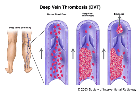
Deep vein thrombosis (DVT) is the formation of a blood clot, known as a thrombus, in the deep leg vein. It is a very serious condition that can cause permanent damage to the leg, known as post-thrombotic syndrome, or a life-threating pulmonary embolism. In the United States alone, 600,000 new cases are diagnosed each year. One in every 100 people who develops DVT dies. Recently, it has been referred to as “Economy Class Syndrome” due to the occurrence after sitting on long flights.
The deep veins that lie near the center of the leg are surrounded by powerful muscles that contract and force deoxygenated blood back to the lungs and heart. One-way valves prevent the back-flow of blood between the contractions. (Blood is squeezed up the leg against gravity and the valves prevent it from flowing back to our feet.) When the circulation of the blood slows down due to illness, injury or inactivity, blood can accumulate or “pool,” which provides an ideal setting for clot formation.
Chronic vs Acute DVT
Chronic DVT refers to venous thrombosis present for more than 28 days. Chronic DVT can either totally permanently block the vein or it can adhere to the wall of the vein. Chronic DVT that doesn’t block the vein can still cause long-term problems as the valves in the vein are often damaged or destroyed. The destruction of the valves can cause the legs to swell after standing for long periods, or cause aching pain and even skin ulcers in severe cases. These symptoms, called post-thrombotic syndrome, are caused by the blood pooling in the legs from gravity.
Acute DVT refers to venous thrombosis for which symptoms have been present for 14 days or less. The symptoms of acute DVT are limb swelling and pain. During this period the clot is soft and easily treated with clot dissolving drugs.
Subacute DVT refers to venous thrombosis that is between acute and chronic. This type of thrombus is starting to form permanent bonds that will eventually turn into a scar like tissue. As the thrombus gets older it shrinks and converts to harder tissue. The longer the thrombus has been “organizing” the tougher it is to dissolve the clot, even with the best drugs. Typically, dissolving the clot is still an option for subacute DVT.
Treatment for DVT
Blood Thinners: Early in treatment, blood thinners are given to keep the clot from growing or breaking off and traveling to the lung. Over time, the body will dissolve most of the clot, but often the vein becomes damaged in the meantime. If the clot were to break off and float to the lung it blocks the blood flow to the lung and is termed “Pulmonary Embolus”. Pulmonary embolus (PE) is a life-threatening complication than can occur from DVT in the leg. The prevention and treatment of PE is discussed under the PE section.
Thrombolysis (Clot Busting Treatment): An invasive procedures to dissolve DVT is performed if a patient has severe pain, difficulty walking, significant swelling or if there is clot blocking the pelvic veins (iliac veins). If someone with a DVT has worsening symptoms while on blood thinners or is having difficulty walking, they should consider evaluation by an Interventional Radiologist. When performed early, thrombolysis is highly effective at dissolving clot and preserving the valves in the veins.
If it is decided that a patient needs clot removed by these methods, it is performed under x-ray guidance by interventional radiologists. This procedure, performed in a hospital’s interventional radiology suite, is designed to rapidly break up the clot, restore blood flow within the vein, and potentially preserve valve function to minimize the risk of post-thrombotic syndrome. The interventional radiologist inserts a tiny tube into the vein behind the knee or other leg vein and threads it into the vein containing the clot using x-ray guidance. The catheter tip is placed into the clot and a “clot busting” drug is infused directly to the thrombus (clot). The fresher the clot, the faster it dissolves – one to two days. Any narrowing in the vein that might lead to future clot formation can be identified during the procedure and treated by the interventional radiologist with a balloon or stent..
Clinical resolution of pain and swelling and restoration of blood flow in the vein is greater than 85 percent with these invasive techniques. In patients in whom thrombolysis or blood thinners are not medically appropriate, an interventional radiologist can insert a vena cava filter, a small device that functions like a umbrella to capture blood clots that would float to the lung, but allows normal liquid blood to pass. Please see that section for more details.
THE SERVICES LISTED ON THIS WEBSITE ARE FOR GENERAL INFORMATION PURPOSES ONLY AND DO NOT INCLUDE ALL SERVICES OF FLORIDA INTERVENTIONAL SPECIALISTS. WHILE WE STRIVE TO KEEP THE INFORMATION UP TO DATE AND CORRECT, WE MAKE NO REPRESENTATIONS OR WARRANTIES OF ANY KIND, EXPRESS OR IMPLIED, ABOUT THE CONTENT, COMPLETENESS, ACCURACY, RELIABILITY, LEGALITY, SUITABILITY OR AVAILABILITY, WITH RESPECT TO THE SERVICES CONTAINED ON THIS WEBSITE.
